
Page 112 - Biology_F5
P. 112
Comparative studies of natural groups of organisms
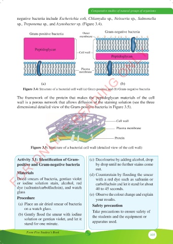
negative bacteria include Escherichia coli, Chlamydia sp., Neisseria sp., Salmonella
sp., Treponema sp., and Azotobacter sp. (Figure 3.4).
Gram-positive bacteria Outer Gram-negative bacteria
membrane
FOR ONLINE READING ONLY
Peptidoglycan
Cell wall
Peptidoglycan
Plasma
membrane
(a) (b)
Figure 3.4: Structure of a bacterial cell wall (a) Gram-positive and (b) Gram-negative bacteria
The framework of the protein that makes the peptidoglycan materials of the cell
wall is a porous network that allows diffusion of the staining solution (see the three
dimensional detailed view of the Gram-positive bacteria in Figure 3.5).
Cell wall
Plasma membrane
Protein
Figure 3.5: Structure of a bacterial cell wall (detailed view of the cell wall)
Activity 3.1: Identification of Gram- (c) Decolourise by adding alcohol, drop
positive and Gram-negative bacteria by drop until no further stains come
out.
Materials (d) Counterstain by flooding the smear
Dried smears of bacteria, gentian violet with a red dye such as safranin or
or iodine solution stain, alcohol, red carbolfuchsin and let it stand for about
dye (safranin/carbolfuchsin), and watch 40 to 45 seconds.
glass (e) Observe the colour change and explain
Procedure your results.
(a) Place an air dried smear of bacteria Safety precaution
on a watch glass.
(b) Gently flood the smear with iodine Take precautions to ensure safety of
the students and the equipment or
solution or gentian violet, and let it
stand for one minute. apparatus used.
Form Five Student’s Book
105