
Page 205 - Biology_F5
P. 205
Biology for Advanced Level Secondary Schools
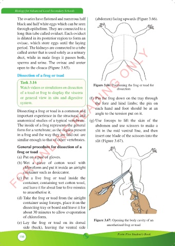
The ovaries have flattened and numerous half (abdomen) facing upwards (Figure 3.66).
black and half white eggs which can be seen
through epithelium. They are connected to a
long thin tube called oviduct. Each oviduct
is dilated in its posterior region to form an
ovisac, which store eggs until the laying
FOR ONLINE READING ONLY
period. The kidneys are connected to a tube
called ureter that is used solely as a urinary
duct, while in male frogs it passes both,
sperms and urine. The ovisac and ureter
open to the cloaca (Figure 3.65).
Dissection of a frog or toad
Task 3.16
Watch videos or simulations on dissection Figure 3.66: Positioning the frog or toad for
dissection
of a toad or frog to display the viscera
or general view in situ and digestive (f) Pin the frog down on the tray through
system. the fore and hind limbs; the pin on
each hand and foot should be at an
Dissecting a frog or toad is a common and angle to the tension put on it.
important experience in the structural and
anatomical studies of a typical vertebrate. (g) Use forceps to lift the skin of the
The inside of a frog represents the general abdomen and use scissors to make a
form for a vertebrate; as the organs present slit in the mid ventral line, and then
in a frog and the way they are laid out are insert one blade of the scissors into the
similar enough to that of other vertebrates. slit (Figure 3.67).
General procedure for dissection of a
frog or toad
(a) Put on a pair of gloves.
(b) Wet a piece of cotton wool with
chloroform and put it inside an airtight
container such as desiccator.
(c) Put a live frog or toad inside the
container, containing wet cotton wool,
and leave it for about four to five minutes
to anaesthetise it.
(d) Take the frog or toad from the airtight
container using forceps, place it on the
dissecting tray or board and leave it for
about 30 minutes to allow evaporation
of chloroform.
(e) Lay the frog or toad on its dorsal Figure 3.67: Opening the body cavity of an
side (back), leaving the ventral side anesthetized frog or toad
Form Five Student’s Book
198