
Page 212 - Biology_F5
P. 212
Comparative studies of natural groups of organisms
(c) The lower jaw is made up of a single immature young ones and duck-billed
bone called the dentary platypus, and echidna which lay eggs.
(d) The middle ear has three small soft bones
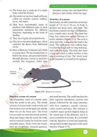
called ear ossicles, namely malleus, Structure of a mouse
incus, and stapes. Structurally, an adult mouse has an average
(e) They have heterotrophic mode of length of 7.5 cm to 10 cm. Its body is
FOR ONLINE READING ONLY
nutrition with different types of teeth divided into head, neck, and trunk. The
(heterodont dentition) for different head bears external ear flaps called pinnae,
functions, depending on the mode of eyes, nostrils and a mouth. The latter has
feeding. long, hair extensions called vibrissae or
(f) They have highly developed brains. tactile whiskers (Figure 3.72). The neck is
(g) They have muscular diaphragm, which short and wide, connecting the head to the
separates the thorax from abdominal trunk. The trunk bears four walking legs,
cavity. two short hind legs and two long front legs,
(h) Most of them are viviparous (give birth each with five digits. The trunk also bears
to young ones). The developing foetus is a long tail which is either hairless or has
held in the uterus, and gets nourishment sparse covering of hair. The whole body
through placenta, except in primitive of the mammal is covered by fur (pelage).
animals like kangaroo, which bear
Hairless ear
Eye
Sensory
whisker
Tail with fur
Hind limb
Short legs clawed feet
Figure 3.72: Structure of a mouse
Digestive system of a mouse and small intestine. The small intestine has
The alimentary canal of a mouse starts three segments (duodenum, jejunum and
from the mouth to the anus. The mouth ileum), followed by the large intestine,
consists of sixteen teeth; twelve molar and with four segments, namely caecum,
four incisors (two on the upper jaw and the colon, rectum and anus. The stomach is
other two on the lower jaw). The lower a hollow organ (pouch-shaped) lying in
incisors teeth are more developed, pointed, the ventral part of the abdomen, and it is
sharp and longer than the restof the teeth, partly covered by liver lobes. It is concerned
and are used for cutting the food into small with the digestion of food and temporary
chewable pieces. Down from the mouth, storage before further digestion in the small
there are oesophagus, stomach, pancreas, intestine (Figure 3.73).
Form Five Student’s Book
205