
Page 36 - Biology_F5
P. 36
Cytology
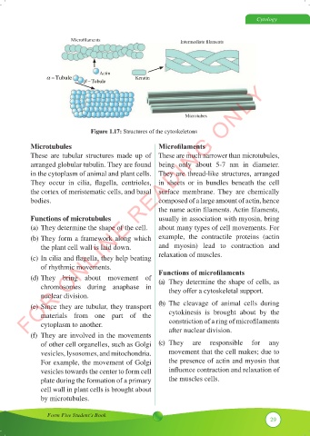
Microfilaments Intermediate filaments
Actin
α −Tubule Keratin
β −Tubule
FOR ONLINE READING ONLY
Microtubes
Figure 1.17: Structures of the cytoskeletons
Microtubules Microfilaments
These are tubular structures made up of These are much narrower than microtubules,
arranged globular tubulin. They are found being only about 5-7 nm in diameter.
in the cytoplasm of animal and plant cells. They are thread-like structures, arranged
They occur in cilia, flagella, centrioles, in sheets or in bundles beneath the cell
the cortex of meristematic cells, and basal surface membrane. They are chemically
bodies. composed of a large amount of actin, hence
the name actin filaments. Actin filaments,
Functions of microtubules usually in association with myosin, bring
(a) They determine the shape of the cell. about many types of cell movements. For
(b) They form a framework along which example, the contractile proteins (actin
the plant cell wall is laid down. and myosin) lead to contraction and
(c) In cilia and flagella, they help beating relaxation of muscles.
of rhythmic movements.
(d) They bring about movement of Functions of microfilaments
(a) They determine the shape of cells, as
chromosomes during anaphase in they offer a cytoskeletal support.
nuclear division.
(e) Since they are tubular, they transport (b) The cleavage of animal cells during
materials from one part of the cytokinesis is brought about by the
cytoplasm to another. constriction of a ring of microfilaments
(f) They are involved in the movements after nuclear division.
of other cell organelles, such as Golgi (c) They are responsible for any
vesicles, lysosomes, and mitochondria. movement that the cell makes; due to
For example, the movement of Golgi the presence of actin and myosin that
vesicles towards the center to form cell influence contraction and relaxation of
plate during the formation of a primary the muscles cells.
cell wall in plant cells is brought about
by microtubules.
Form Five Student’s Book
29