
Page 85 - Biology Form Two
P. 85
Biology for Secondary Schools
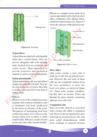
Phloem is a complex tissue made up of
Pits various specialised cells which are sieve
tubes, companion cells, phloem fibres,
and phloem parenchyma cells. Figure 4.5
shows the structure of the phloem tissue.
FOR ONLINE READING ONLY
Tapering end Nucleus
Companion cell
Sieve plate
Plasmodesmata
Figure 4.4: Tracheids
Mitochondria
Xylem fibres
Xylem fibres are dead cells with lignified
walls and a central lumen. They are Sieve tube elements
narrow, elongated cells with tapering
ends, located between tracheids and Figure 4.5: Phloem tissue
xylem vessels. Their function is to
provide protection and mechanical Sieve tubes
support to xylem vessels and tracheids. Like xylem vessels, a sieve tube is
made up of cells that are joined end-to-
Xylem parenchyma end. However, the end walls of these
Xylem parenchyma are non-specialised cells are not completely broken down
simple cells with thin cell walls. They are but have perforations or pores that
the only living cells of xylem. They help form sieve plates, as shown in Figure
in storing water and food in the form of 4.5. These cells contain cytoplasm,
carbohydrate.
but they have no nucleus. Fibres run
Phloem through the pores, thereby connecting
Phloem is one of the conducting tissues adjacent sieve tube cells.
in plants that conducts substances such
as hormones and food synthesised Companion cells
in the leaves to all parts of the plant. Each sieve tube element is associated
The process of transporting food from with a living cell called a companion
synthesising organs such as leaves to the cell. The two cells are separated by a thin
storage organs such as tubers is called wall made up of parenchyma cells with
translocation. Phloem is usually located pores called plasmodesmata, which
on the outer side of the vascular bundles. allow exchange of materials between
Student’s Book - Form Two 79
05/10/2024 15:36:28
BIOLOGY FORM 2 NEW.indd 79 05/10/2024 15:36:28
BIOLOGY FORM 2 NEW.indd 79