Page 248 - Biology_F5
P. 248
Coordination and Irritability

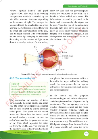
           cornea,  aqueous  humour  and  pupil  there  are  cone  and  rod  photoreceptors,
           (Figure 4.30).  The pupil is an opening  which are connected to the brain via a
           (an aperture), which is controlled by the  bundle  of  fibres  called  optic  nerve.  The
           iris  (like  camera  shutters)  depending  information  received  is  processed  in  the
           on the amount of light. The stronger the  brain, and consequently, the object can
           amount of light, the smaller the size of the  be seen. Thus, the role of the retina is to
          FOR ONLINE READING ONLY
           aperture is. The lens is positioned between  translate  light  into  nerve signals and to
           the outer and inner chambers of the eye,  allow us to see under various conditions
           and its major function is to focus images  ranging from starlight to sunlight. It also
           on the  retina  by changing  its  thickness  distinguishes  the  wavelengths for us to
           depending  on the  amount  of light  from  discriminate colors.
           distant  or nearby objects.  On the  retina,

                                                      Light
                                     Iris

                                                                  Retina

                                                                  Inverted image
                                                                  of object


                          Object

                                           Lens            Optic nerves

                       Figure 4.30: Structure of a mammalian eye showing physiology of seeing
           4.2.3  The mammalian ear                   and glands that secrete earwax, which is
                                                      located in the upper wall of the auditory
             Task 4.9                                 canal.  Ear wax guards the ear against
             Search from the internet sources on the   entrance of foreign materials such as dust
             simulations or videos on the mechanism   and microorganisms.
             of hearing and body balance make short
             notes on the searched information        In the middle  ear, the  tympanic
                                                      membrane begins and ends at a bony wall
           The  mammalian  ear consists of three      containing two small openings covered by
           parts, namely the outer, middle and inner   membranes.  The two openings are oval
           ear. The outer ear comprises an external   window (fenestra  ovalis)  and the  round
           flap  of  skin  covered  by  elastic  cartilage   window (fenestra rotunda).  There are
           called  pinna.  The  pinna  collects  sound   three connected bones called ear ossicles,
           waves and directs them into the ear canal   which are held in position by muscles.
           (external  auditory  meatus).  Across the   These are malleus (hammer), incus (anvil),
           end of ear canal is a tympanic membrane    and stapes (stirrup). The middle ear is air
           or  ear  drum  which  separates  the  outer   filled part that depends on the equalization
           ear from the middle ear. The opening of    of pressure outside and inside the ear to
           the auditory canal is lined with fine hairs

                 Form Five Student’s Book
                                                                                          241

