
Page 250 - Biology_F5
P. 250
Coordination and Irritability
Semicircular canals
Utricle
Saccule
FOR ONLINE READING ONLY
Cochlea
Endolymphatic duct
Figure 4.32: Components of the membranous labyrinth
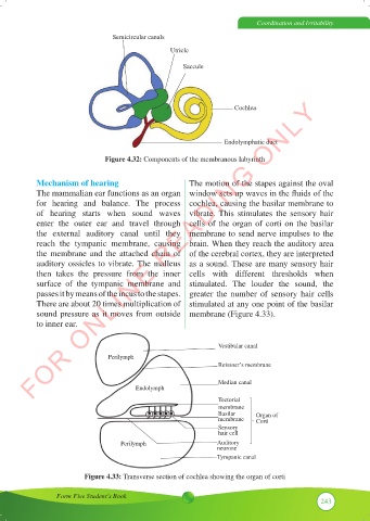
Mechanism of hearing The motion of the stapes against the oval
The mammalian ear functions as an organ window sets up waves in the fluids of the
for hearing and balance. The process cochlea, causing the basilar membrane to
of hearing starts when sound waves vibrate. This stimulates the sensory hair
enter the outer ear and travel through cells of the organ of corti on the basilar
the external auditory canal until they membrane to send nerve impulses to the
reach the tympanic membrane, causing brain. When they reach the auditory area
the membrane and the attached chain of of the cerebral cortex, they are interpreted
auditory ossicles to vibrate. The malleus as a sound. These are many sensory hair
then takes the pressure from the inner cells with different thresholds when
surface of the tympanic membrane and stimulated. The louder the sound, the
passes it by means of the incus to the stapes. greater the number of sensory hair cells
There are about 20 times multiplication of stimulated at any one point of the basilar
sound pressure as it moves from outside membrane (Figure 4.33).
to inner ear.
Vestibular canal
Perilymph
Reissner’s membrane
Median canal
Endolymph
Tectorial
membrane
Basilar Organ of
membrane Corti
Sensory
hair cell
Perilymph Auditory
neurone
Tympanic canal
Figure 4.33: Transverse section of cochlea showing the organ of corti
Form Five Student’s Book
243