
Page 251 - Biology_F5
P. 251
Biology for Advanced Level Secondary Schools
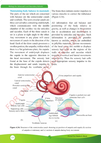
Maintaining body balance in mammals The brain then initiates motor impulses to
The parts of the ear which are concerned various muscles to correct the imbalance
with balance are the semicircular canals (Figure 4.34).
and vestibule. The semi circular canals are
three curved tubes containing endolymph, All information that aid balance and
which communicates with the middle positioning of the body relative to
FOR ONLINE READING ONLY
chamber of the cochlea via the utriculus gravity, as well as changes in the position
and sacculus. Each of the three canals is due to acceleration and deceleration is
set in a plane at right angle to the other. provided by utriculus and sacculus. Such
Any movement in any plane will cause information is provided by granules
movement of canals in the direction of the known as otoliths, which are embedded
head. Each of the three canals possesses a in jellylike materials. Various movements
swollen portion, the ampulla, within which of the head cause this otolith to displace
there is a flat gelatinous plate, the cupula. sensory hair cells on the regions of the
The movement of endolymph displaces walls of utriculus and sacculus which
the cupula in the opposite direction to respond to vertical and lateral movements
the head movement. The sensory hair respectively. Then the sensory hair cells
found at the base of the cupula detects send appropriate sensory impulse to the
the displacement and sends impulse to brain.
the brain through the vestibular nerve.
Anterior semicircular Crista ampullaris and cupula
canal
Lateral semicircular
canal
Posterior semicircular
canal
Ampullae
(a) Cupula is pushed over and
Crista Direction stimulates hair cells
Cupula ampullaris of rotation
Endolymph
Sensory hair cells
Supporting cells
Sensory nerve fiber
Endolymph
lags behind Stereocilia are bent
due to inertia
(b) (c)
Vestibular neurone
Figure 4.34: Structure of the semicircular canals showing (a) position of ampula and cupula (b) section
of ampula at stationary and (c) section of ampula during body movement
Form Five Student’s Book
244