
Page 300 - Biology_F5
P. 300
Gas exchange and respiration
The diaphragm separates the thorax from the and goblet cells. The cilia beating moves the
abdomen. During inhalation the volume of trapped dusts and bacteria back to the cavity
the thoracic cavity increases, thus lowering where they get swallowed. The goblet cells
pressure. This is caused by the downward are essential for mucus production that traps
movement of the diaphragm and the outward dusts and bacteria altogether. It also moistens
movement of the ribs. During exhalation, the air that passes down to the alveoli. The
FOR ONLINE READING ONLY
the volume of the thoracic cavity decreases, trachea branches at its lower end into two
thus raising the pressure. This is caused by bronchi. Like the trachea, each bronchus
the upward movement of the diaphragm and has cartilage and ciliated epithelium with
inward movement of the ribs. goblet cells which play a role of trapping dust
and bacteria. Each bronchus subdivides into
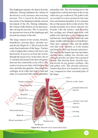
The lungs consist of the trachea, bronchi, many smaller tubes known as bronchioles.
bronchioles, alveolar ducts, alveolar sacs They have cartilage, cilia, and the goblet
and alveoli (Figure 6.1). The alveoli are the cells that work similarly as in the trachea
main functional units of the lungs. Trachea and bronchi; they trap foreign substances.
is the windpipe that connects the larynx and The bronchioles branch into very fine tubes,
the bronchi. It receives air from the nostrils alveolar ducts, ending into alveolar sacs
through the nasal cavity. In the latter, the air and each alveolar sac opens into a group of
is warmed and cleared from dust and germs alveoli. The alveolar ducts, alveolar sacs,
that may have entered the cavity with it. The and alveoli do not possess cartilage, cilia
nasal cavity possesses hairs which help to trap and goblet cells. The alveolus possesses
dusts and other tiny particles. The trachea has features that allow it to function in a special
cartilages in its walls that keep it firm and way compared to other structural parts of
intact. It is associated with ciliated epithelium the lungs.
Larynx
Trachea (windpipe) Clavicle (collar bone)
Ring of cartilage
External intercostal
Right lung muscles
Bronchus
Bronchioles
Internal intercostal
Alveoli muscles
Sternum
Intercostal muscle
Left lung
Right rib Left rib
Pleural membrane Pleural cavity
Diaphragm
Figure 6.1: Structure of the mammalian lungs
Form Five Student’s Book
293