
Page 235 - Physics_Form_2
P. 235
Optical instruments
Structure of the human eye
holding a camera. If you wish to
take a picture of your own image, The eye is nearly spherical and is about
at what distance should you focus 2.5 cm in diameter. The front portion is
your camera? somehow more sharply curved and is
3. A lens camera is used to photograph a covered by a tough, transparent membrane
FOR ONLINE READING ONLY
called the cornea. The region behind the
person who is 2.8 m tall and standing
2.7 m in front of the camera. If the cornea contains a liquid called aqueous
humour. Next to the aqueous humour,
film is placed 10 cm behind the lens,
calculate the height of the produced there is a crystalline lens, which is a
capsule containing a fibrous jelly, hard at
image.
the centre and progressively softer at the
outer portions. The crystalline lens is held
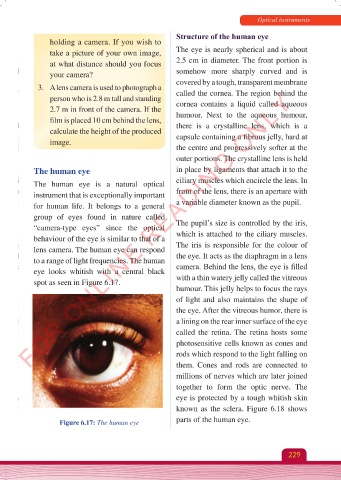
The human eye in place by ligaments that attach it to the
The human eye is a natural optical ciliary muscles which encircle the lens. In
instrument that is exceptionally important front of the lens, there is an aperture with
for human life. It belongs to a general a variable diameter known as the pupil.
group of eyes found in nature called
“camera-type eyes” since the optical The pupil’s size is controlled by the iris,
behaviour of the eye is similar to that of a which is attached to the ciliary muscles.
lens camera. The human eye can respond The iris is responsible for the colour of
to a range of light frequencies. The human the eye. It acts as the diaphragm in a lens
eye looks whitish with a central black camera. Behind the lens, the eye is filled
spot as seen in Figure 6.17. with a thin watery jelly called the vitreous
humour. This jelly helps to focus the rays
of light and also maintains the shape of
the eye. After the vitreous humor, there is
a lining on the rear inner surface of the eye
called the retina. The retina hosts some
photosensitive cells known as cones and
rods which respond to the light falling on
them. Cones and rods are connected to
millions of nerves which are later joined
together to form the optic nerve. The
eye is protected by a tough whitish skin
known as the sclera. Figure 6.18 shows
Figure 6.17: The human eye parts of the human eye.
229
Physics Form 2 Final.indd 229 25/10/2025 10:28