
Page 55 - Biology_Form_3
P. 55
Coordination in animals
Structurally the eye is made up of three The middle layer is choroid; this layer is
layers of tissues which are called sclerotic highly pigmented. It is a nutritive layer
layers or sclera, choroid and retina. The which is located just next to sclera. The
outermost layer of the eye is extremely layer extends to the front part of the eye
tough and it is called the sclera. This layer where it forms ciliary body and iris. The
FOR ONLINE READING ONLY
protects, supports, and maintains the shape choroid also contains a network of blood
of the eyeball. The sclera is white in colour vessels which nourish and supply oxygen
except the front part which is transparent to the eye. This layer has black coloured
and it is called cornea. It also contains pigments on its wall. All the light entering
elastic connective tissues. The cornea is the eye is absorbed by the black pigment
the transparent part of the eyeball which is in the choroid.
continuous with the sclera and is covered
with a thin membrane called conjunctiva. Iris is a ring of contractile muscles which
Cornea is convex so that light rays can is an extension of the ciliary body. This
be refracted, and since it is transparent, it part is made up of two sets of muscles
allows light to pass through. called circular and radial muscles. These
muscles are able to contract and relax. The
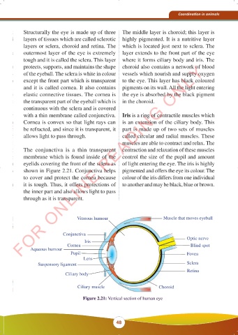
The conjunctiva is a thin transparent contraction and relaxation of these muscles
membrane which is found inside of the control the size of the pupil and amount
eyelids covering the front of the sclera as of light entering the eye. The iris is highly
shown in Figure 2.21. Conjunctiva helps pigmented and offers the eye its colour. The
to cover and protect the cornea because colour of the iris differs from one individual
it is tough. Thus, it offers protections of to another and may be black, blue or brown.
the inner part and also allows light to pass
through as it is transparent.
Vitreous humour Muscle that moves eyeball
Conjunctiva
Iris Optic nerve
Cornea Blind spot
Aqueous humour
Pupil Fovea
Lens
Suspensory ligament Sclera
Retina
Ciliary body
Ciliary muscle Choroid
Figure 2.21: Vertical section of human eye
48
23/10/2025 11:52:37
BIOLOGY FORM 3 FINAL EDITED 16.09.2025.indd 48
BIOLOGY FORM 3 FINAL EDITED 16.09.2025.indd 48 KAMISHNA 23/10/2025 11:52:37
KAMISHNA