
Page 50 - Biology_Form_3
P. 50
Biology for Secondary Schools
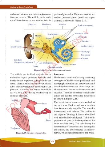
and round window which is also known as position by muscles. These ear ossicles are
fenestra rotunda. The middle ear is made malleus (hammer), incus (anvil) and stapes
up of three bones or ear ossicles held in (stirrup) as shown in Figure 2.16.
Outer ear Middle ear Inner ear Semicircular canals
FOR ONLINE READING ONLY
Oval window Vestibular nerve
Malleus Utriculus
Stapes
Pinna External Sacculus
ear canal
Sound waves Auditory nerve
Endolymph
Perilymph
Cochlea
Ear drum
(Tympanic membrane) Air filled space
Round window
Incus Eustachian tube
Figure 2.16: Structure of the mammalian ear
The middle ear is filled with air which Inner ear
maintains equal pressure outside and The inner ear consists of a cavity containing
inside the ear to prevent damage to the ear two types of fluids called perilymph and
drum. There is a Eustachian tube (auditory endolymph. It consists of a membranous and
tube) which connects the middle ear to the bony labyrinth composed of two large sac-
pharynx. Air enters and leaves the middle like structures, known as the utriculus and
ear via this tube during swallowing to sacculus. There are also three semicircular
equalize pressure. canals and a coiled tube called the cochlea
as shown in Figure 2.16.
Malleus
Incus The semicircular canals are attached to
the utriculus. Each canal has a swollen
base known as the ampulla. The ampulla
is concerned with balance. The cochlea is
the organ of hearing. It has a tube filled
with a fluid called endolymph. This fluid is
Stapes present in all parts of the bony tubes of the
inner ear (labyrinth). The cells lining the
inner surface of the cochlea and the ampulla
Tympanum
are sensory and are connected to auditory
Figure 2.17: Structure of middle Ear nerves, which send impulses to the brain.
43 Student’s Book - Form Three
23/10/2025 11:52:36
BIOLOGY FORM 3 FINAL EDITED 16.09.2025.indd 43
BIOLOGY FORM 3 FINAL EDITED 16.09.2025.indd 43 KAMISHNA 23/10/2025 11:52:36 KAMISHNA