
Page 92 - Biology_Form_3
P. 92
Excretion
The ureters before being discharged out of the body. At
These are two tubes of smooth muscle that the base of the urinary bladder there is a
extend from the pelvic region of the kidney sphincter muscle that prevents the bladder
to the urinary bladder. Their major function from emptying the urine until it reaches a
is to carry urine from the kidney to the certain level.
FOR ONLINE READING ONLY
urinary bladder. The entrance to the bladder
has a ureterovesical valve that prevents the The urethra
urine from flowing back to the kidney. This refers to a thin, fibromuscular tube that
begins at the lower opening of the bladder
The urinary bladder and extends through the pelvis to the outside
This is a spherical muscular sac that is of the body see Figure 4.1. The wall of the
located within the pelvic cavity. The waste urethra is composed of mucous membrane
fluid that is produced in the liver as a result and fibrous smooth muscle tissue. These
of metabolic processes is transported in the are the muscles that release the urine to the
bloodstream to the kidney where the waste outside of the body. In males, the ductus
products are filtered out and become part deferens connects to the uretha to deliver
of the urine. The urine is transferred into the sperms during ejaculation.
urinary bladder where it is temporarily stored
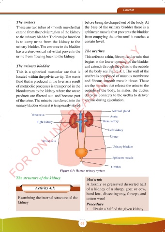
Adrenal gland
Vena cava Aorta
Right kidney Renal artery
Left kidney
Ureter
Renal vein
Urinary bladder
Sphincter muscle
Urethra
Figure 4.1: Human urinary system
The structure of the kidney Materials
A freshly or preserved dissected half
Activity 4.1: of a kidney of a sheep, goat or cow,
hand lens, dissecting tray, forceps, and
Examining the internal structure of the cotton wool
kidney Procedure
1. Obtain a half of the given kidney.
85
23/10/2025 11:52:43
BIOLOGY FORM 3 FINAL EDITED 16.09.2025.indd 85
BIOLOGY FORM 3 FINAL EDITED 16.09.2025.indd 85 KAMISHNA 23/10/2025 11:52:43
KAMISHNA