
Page 105 - Biology Form Two
P. 105
Biology for Secondary Schools
expanding when it is beating very fast. gets fatigued; they work continuously
The pericardium also protects the heart as long as a person is alive. This type
from mechanical injury and friction. of muscle is found only in the heart.
It provides enough room for vigorous The inner layer is called endocardium.
pumping of the heart. In addition, the
FOR ONLINE READING ONLY
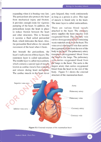
pericardium keeps the heart in place There are various blood vessels
to reduce friction between the heart attached to the heart. The coronary
and other structures. This is because artery supplies the heart muscles with
it secretes a fluid called pericardial oxygenated blood. The coronary vein
fluid, which lubricates the heart. Also, carries deoxygenated blood containing
the pericardial fluid allows for smooth waste materials away from the heart. The
vena cava is the largest vein that carries
movement of the heart when it beats.
deoxygenated blood from the rest of the
Just beneath the pericardium, the body to the heart. The pulmonary artery
heart’s wall consists of three layers. The transports deoxygenated blood from
outermost layer is called epicardium. the heart to the lungs. The pulmonary
The middle layer is called myocardium, vein carries oxygenated blood from
which contains a special type of muscle the lungs to the heart. The aorta is the
known as cardiac muscle that contracts largest artery that carries oxygenated
and relaxes during heart movement. blood from the heart to the rest of the
The cardiac muscle in the heart never body. Figure 5.1 shows the external
structure of the mammalian heart.
Superior vena cava
Aorta
Pulmonary artery
Pulmonary veins
Inferior vena cava Coronary artery
Coronary vein
Figure 5.1: External structure of the mammalian heart
Student’s Book - Form Two
99
05/10/2024 15:36:37
BIOLOGY FORM 2 NEW.indd 99
BIOLOGY FORM 2 NEW.indd 99 05/10/2024 15:36:37